Review


human 130 125 042  (Miltenyi Biotec)


Bioz Verified Symbol Miltenyi Biotec is a verified supplier
Bioz Manufacturer Symbol Miltenyi Biotec manufactures this product  
  • Logo
  • About
  • News
  • Press Release
  • Team
  • Advisors
  • Partners
  • Contact
  • Bioz Stars
  • Bioz vStars
  • 93

    Structured Review

    Miltenyi Biotec human 130 125 042
    Human 130 125 042, supplied by Miltenyi Biotec, used in various techniques. Bioz Stars score: 93/100, based on 5 PubMed citations. ZERO BIAS - scores, article reviews, protocol conditions and more
    https://www.bioz.com/product/human+130+125+042/StemMACS+HSC-CFU+Assay+Kit%2C+human/pmc12858490-44-45-30
    Average 93 stars, based on 5 article reviews
    human 130 125 042 - by Bioz Stars, 2026-09
    93/100 stars

    Images


    Similar Products

    93
    Miltenyi Biotec human 130 125 042
    Human 130 125 042, supplied by Miltenyi Biotec, used in various techniques. Bioz Stars score: 93/100, based on 1 PubMed citations. ZERO BIAS - scores, article reviews, protocol conditions and more
    https://www.bioz.com/product/human+130+125+042/StemMACS+HSC-CFU+Assay+Kit%2C+human/pmc12858490-44-45-30
    Average 93 stars, based on 1 article reviews
    human 130 125 042 - by Bioz Stars, 2026-09
    93/100 stars
      Buy from Supplier

    93
    Miltenyi Biotec assay kit
    Assay Kit, supplied by Miltenyi Biotec, used in various techniques. Bioz Stars score: 93/100, based on 1 PubMed citations. ZERO BIAS - scores, article reviews, protocol conditions and more
    https://www.bioz.com/product/human+130+125+042/StemMACS+HSC-CFU+Assay+Kit%2C+human/pmc12858490-44-24-30
    Average 93 stars, based on 1 article reviews
    assay kit - by Bioz Stars, 2026-09
    93/100 stars
      Buy from Supplier

    93
    Miltenyi Biotec lineage potential
    Lineage Potential, supplied by Miltenyi Biotec, used in various techniques. Bioz Stars score: 93/100, based on 1 PubMed citations. ZERO BIAS - scores, article reviews, protocol conditions and more
    https://www.bioz.com/product/human+130+125+042/StemMACS+HSC-CFU+Assay+Kit%2C+human/pmc12858490-44-9-30
    Average 93 stars, based on 1 article reviews
    lineage potential - by Bioz Stars, 2026-09
    93/100 stars
      Buy from Supplier

    93
    Miltenyi Biotec stemmacs hsccfu assay cocktail
    Stemmacs Hsccfu Assay Cocktail, supplied by Miltenyi Biotec, used in various techniques. Bioz Stars score: 93/100, based on 1 PubMed citations. ZERO BIAS - scores, article reviews, protocol conditions and more
    https://www.bioz.com/product/human+130+125+042/StemMACS+HSC-CFU+Assay+Kit%2C+human/pm41452425-50-9-15
    Average 93 stars, based on 1 article reviews
    stemmacs hsccfu assay cocktail - by Bioz Stars, 2026-09
    93/100 stars
      Buy from Supplier

    93
    Miltenyi Biotec assay cocktail
    Assay Cocktail, supplied by Miltenyi Biotec, used in various techniques. Bioz Stars score: 93/100, based on 1 PubMed citations. ZERO BIAS - scores, article reviews, protocol conditions and more
    https://www.bioz.com/product/human+130+125+042/StemMACS+HSC-CFU+Assay+Kit%2C+human/pmc12858490-60-11-15
    Average 93 stars, based on 1 article reviews
    assay cocktail - by Bioz Stars, 2026-09
    93/100 stars
      Buy from Supplier

    93
    Miltenyi Biotec methylcellulose based medium
    Methylcellulose Based Medium, supplied by Miltenyi Biotec, used in various techniques. Bioz Stars score: 93/100, based on 1 PubMed citations. ZERO BIAS - scores, article reviews, protocol conditions and more
    https://www.bioz.com/product/human+130+125+042/StemMACS+HSC-CFU+Assay+Kit%2C+human/pmc12012840__HEM3___9___e70120___s003-58-21-28
    Average 93 stars, based on 1 article reviews
    methylcellulose based medium - by Bioz Stars, 2026-09
    93/100 stars
      Buy from Supplier

    93
    Miltenyi Biotec medium
    Medium, supplied by Miltenyi Biotec, used in various techniques. Bioz Stars score: 93/100, based on 1 PubMed citations. ZERO BIAS - scores, article reviews, protocol conditions and more
    https://www.bioz.com/product/human+130+125+042/StemMACS+HSC-CFU+Assay+Kit%2C+human/bio_rxiv__2024__04__08__588553-236-23-29
    Average 93 stars, based on 1 article reviews
    medium - by Bioz Stars, 2026-09
    93/100 stars
      Buy from Supplier

    93
    Miltenyi Biotec human cd34 cells
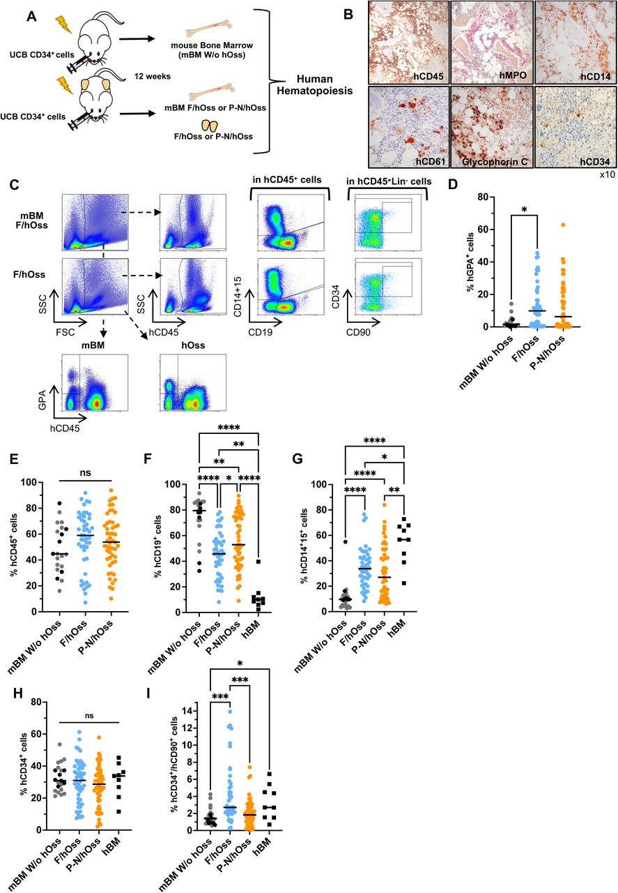
    A. Experimental design of xenografts of umbilical cord blood <t>CD34</t> + cells in mice carrying hOss. Umbilical cord blood CD34 + cells (10 5 cells/mouse) were injected into mice carrying hOss and non-hOss controls, less than 24 hours after sublethal irradiation, 2-8 weeks after hOss implantation. Human hematopoiesis was analyzed 12 weeks after umbilical cord blood HSPC injection into the bone marrow of mice (mBM) and the hOss. B. Umbilical cord blood CD34 + injected in F/hOss at 12 weeks with histology immunolabeling of sections: human hematopoietic cells (hCD45), myeloid cells (hMPO, CD14), megakaryocytes (CD61), red cells (Glycophorin C), immature HSPCs (CD34). Amplification, ×10. C. A representative flow cytometry analysis of the human hematopoietic cells developing in mBM (upper panel) and F/hOss (lower panel). Analysis at 12 weeks post-injection of umbilical cord blood CD34 + cells. D. Percentage of human GPA + cells in mice with and without hOss. Cells were gated on FSC/SSC following the gating strategy in C. Each dot represents individual mice (n=21 mice, without hOss in black and gray from 2 independent experiments; n=25 mice, with F/hOss, 47 hOss obtained from 5 different MSCs; n=28 mice with P-N/hOss, 51 hOss obtained from 4 different MSCs). Gray and black dots: mBM from mice without hOss; blue dots: F/hOss; orange dots: P-N/hOss. E. Percentage of cells expressing hCD45 + using the gating strategy shown in C for mBM (pooled 4 long bones) and hOss (2/mouse, treated separately) 12 weeks post-transplant of umbilical cord blood CD34 cells. The results represent the same mice as those displayed in D. F-G. Percentage of human (F) lymphoid CD19 + B-cells and (G) myeloid CD14 + /CD15 cells. Cells were gated on hCD45 + as described in C. hBM cells from healthy donors (age <18 years, n=9) were included as controls. The results represent the same mice as those displayed in D-E. The gating strategy is shown in SupFig 2D and . H-I. Percentage of CD34 + (H) and CD34 + CD90 + HSCs (I) gated on hCD45 + Lin (CD19 CD14 CD15) cells, based on the gating strategy described in C. hBM cells from healthy donors (age <18 years, n=9) were included as controls. The results represent the same mice as those displayed in D-E. ns, no statistical difference; *, p<0.05; **, p<0.001; ****, p<0.0001; Kruskal-Wallis test without correction.
    Human Cd34 Cells, supplied by Miltenyi Biotec, used in various techniques. Bioz Stars score: 93/100, based on 1 PubMed citations. ZERO BIAS - scores, article reviews, protocol conditions and more
    https://www.bioz.com/product/human+130+125+042/StemMACS+HSC-CFU+Assay+Kit%2C+human/bio_rxiv__2024__04__08__588553-236-8-29
    Average 93 stars, based on 1 article reviews
    human cd34 cells - by Bioz Stars, 2026-09
    93/100 stars
      Buy from Supplier

    Image Search Results


    A. Experimental design of xenografts of umbilical cord blood CD34 + cells in mice carrying hOss. Umbilical cord blood CD34 + cells (10 5 cells/mouse) were injected into mice carrying hOss and non-hOss controls, less than 24 hours after sublethal irradiation, 2-8 weeks after hOss implantation. Human hematopoiesis was analyzed 12 weeks after umbilical cord blood HSPC injection into the bone marrow of mice (mBM) and the hOss. B. Umbilical cord blood CD34 + injected in F/hOss at 12 weeks with histology immunolabeling of sections: human hematopoietic cells (hCD45), myeloid cells (hMPO, CD14), megakaryocytes (CD61), red cells (Glycophorin C), immature HSPCs (CD34). Amplification, ×10. C. A representative flow cytometry analysis of the human hematopoietic cells developing in mBM (upper panel) and F/hOss (lower panel). Analysis at 12 weeks post-injection of umbilical cord blood CD34 + cells. D. Percentage of human GPA + cells in mice with and without hOss. Cells were gated on FSC/SSC following the gating strategy in C. Each dot represents individual mice (n=21 mice, without hOss in black and gray from 2 independent experiments; n=25 mice, with F/hOss, 47 hOss obtained from 5 different MSCs; n=28 mice with P-N/hOss, 51 hOss obtained from 4 different MSCs). Gray and black dots: mBM from mice without hOss; blue dots: F/hOss; orange dots: P-N/hOss. E. Percentage of cells expressing hCD45 + using the gating strategy shown in C for mBM (pooled 4 long bones) and hOss (2/mouse, treated separately) 12 weeks post-transplant of umbilical cord blood CD34 cells. The results represent the same mice as those displayed in D. F-G. Percentage of human (F) lymphoid CD19 + B-cells and (G) myeloid CD14 + /CD15 cells. Cells were gated on hCD45 + as described in C. hBM cells from healthy donors (age <18 years, n=9) were included as controls. The results represent the same mice as those displayed in D-E. The gating strategy is shown in SupFig 2D and . H-I. Percentage of CD34 + (H) and CD34 + CD90 + HSCs (I) gated on hCD45 + Lin (CD19 CD14 CD15) cells, based on the gating strategy described in C. hBM cells from healthy donors (age <18 years, n=9) were included as controls. The results represent the same mice as those displayed in D-E. ns, no statistical difference; *, p<0.05; **, p<0.001; ****, p<0.0001; Kruskal-Wallis test without correction.

    Journal: bioRxiv

    Article Title: Humanized in vivo bone marrow models orchestrate multi-lineage human hematopoietic cell development

    doi: 10.1101/2024.04.08.588553

    Figure Lengend Snippet: A. Experimental design of xenografts of umbilical cord blood CD34 + cells in mice carrying hOss. Umbilical cord blood CD34 + cells (10 5 cells/mouse) were injected into mice carrying hOss and non-hOss controls, less than 24 hours after sublethal irradiation, 2-8 weeks after hOss implantation. Human hematopoiesis was analyzed 12 weeks after umbilical cord blood HSPC injection into the bone marrow of mice (mBM) and the hOss. B. Umbilical cord blood CD34 + injected in F/hOss at 12 weeks with histology immunolabeling of sections: human hematopoietic cells (hCD45), myeloid cells (hMPO, CD14), megakaryocytes (CD61), red cells (Glycophorin C), immature HSPCs (CD34). Amplification, ×10. C. A representative flow cytometry analysis of the human hematopoietic cells developing in mBM (upper panel) and F/hOss (lower panel). Analysis at 12 weeks post-injection of umbilical cord blood CD34 + cells. D. Percentage of human GPA + cells in mice with and without hOss. Cells were gated on FSC/SSC following the gating strategy in C. Each dot represents individual mice (n=21 mice, without hOss in black and gray from 2 independent experiments; n=25 mice, with F/hOss, 47 hOss obtained from 5 different MSCs; n=28 mice with P-N/hOss, 51 hOss obtained from 4 different MSCs). Gray and black dots: mBM from mice without hOss; blue dots: F/hOss; orange dots: P-N/hOss. E. Percentage of cells expressing hCD45 + using the gating strategy shown in C for mBM (pooled 4 long bones) and hOss (2/mouse, treated separately) 12 weeks post-transplant of umbilical cord blood CD34 cells. The results represent the same mice as those displayed in D. F-G. Percentage of human (F) lymphoid CD19 + B-cells and (G) myeloid CD14 + /CD15 cells. Cells were gated on hCD45 + as described in C. hBM cells from healthy donors (age <18 years, n=9) were included as controls. The results represent the same mice as those displayed in D-E. The gating strategy is shown in SupFig 2D and . H-I. Percentage of CD34 + (H) and CD34 + CD90 + HSCs (I) gated on hCD45 + Lin (CD19 CD14 CD15) cells, based on the gating strategy described in C. hBM cells from healthy donors (age <18 years, n=9) were included as controls. The results represent the same mice as those displayed in D-E. ns, no statistical difference; *, p<0.05; **, p<0.001; ****, p<0.0001; Kruskal-Wallis test without correction.

    Article Snippet: Based on these results, an equivalent of 1,000 human CD34 + cells from mBM and hOss were plated in 1 mL of methylcellulose-based medium (130-125-042, StemMACSTM HSC-CFU Assay Kit; Miltenyi Biotec) that contained human SCF, IL-3, IL-6, Erythropoietin (EPO), G-CSF, GM-CSF and 1% Penicillin/Streptomycin.

    Techniques: Injection, Irradiation, Immunolabeling, Amplification, Flow Cytometry, Expressing

    A. Experimental design of secondary hOss formation and grafts of CD34 + cells in mice carrying secondary hOss. Secondary hMSCs were isolated after crushing primary hOss, expanded as adherent cells and transplanted as indicated in . Eight weeks later, 10 5 umbilical cord blood CD34 + cells were injected into mice carrying secondary hOss and non-hOss controls less than 24 hours after sublethal (2 Gy/mouse) irradiation. Human hematopoietic development was analyzed 12 weeks later in the bone marrow (BM) of mice and hOss. B. Flow cytometry analysis of secondary hMSCs isolated from primary F/ and P-N/hOss. Cells were gated on hCD45 - CD14 - CD15 - CD31 - cells (>90%). Representative of >4 tested secondary hMSC samples. The results were obtained from the gating strategy shown in SupFig1A. C. Representative examples of secondary hOss obtained 10-12 weeks post-injection of secondary hMSCs. The images display the results for the samples F28 (fetal hMSC origin, 11.6 PCWs) and ALLO3 (post-natal hMSC origin, 5 years old). D. Weight comparison of secondary F/hOss and P-N/hOss. The results were obtained from 18 hOss (individual blue dots, median value: black line) generated from secondary F/hMSCs, derived from 2 MSCs samples, in 3 independent experiments, and obtained from 8 hOss (individual red dots, median value: black line) generated from secondary P-N/hMSCs, derived from 1 hMSC sample in 2 independent experiments. E. Percentage of murine cells analyzed by flow cytometry, recovered from secondary hOss without injection of umbilical cord blood CD34 + cells. Gating was carried out on viable murine red blood cells, Ter119 + , and viable murine hematopoietic cells (in Ter119 - ), mCD45 + . The results are shown for 12 hOss generated with 1 secondary F/hMSC in 2 independent experiments and 2 hOss from 1 secondary P-N/hMSC in 1 experiment. F. Percentage of human GPA + erythroid cells in mBM from mice with and without secondary (II) hOss. The gating strategy is the same as in . The results shown represent the individual values of mBM from 21 mice without hOss (gray dots: the same mice as displayed in ), 8 hOss generated by 2 secondary F/hMSCs in 4 mice (dark blue dots), 6 hOss generated by 1 secondary P-N/hMSC in 3 mice (dark red dots), 6 mBM from mice with secondary F/hOss (light blue dots) and 3 mBM from the mice with secondary P-N/hOss (pink dots). Black lines indicate the median values. G. Percentage of hCD45 + cells assessed via flow cytometry in secondary hOss at 12 weeks post-transplant of umbilical cord blood CD34 + cells. The results represent the same mice as those displayed in F. The gating strategy is the same as that shown in . H-I. Percentage of human (H) lymphoid B-cells (CD19 + ) and (I) myeloid cells (CD14 + /CD15 + ) gated on hCD45+ cells according to the strategy shown in . The results represent the same mice as those displayed in F-G. J-K. Percentage of (J) CD34 + and (K) CD34 + CD90 + HSCs gated on hCD45 + Lin - (CD19 - CD14 - CD15 - ) cells, based on the same gating strategy as that shown in . The results represent the same mice as those displayed in F-G. *, p<0.05; **, p<0.01; ***, p<0.001; ****, p<0.0001; F-K. Kruskal-Wallis test without correction. D: Mann-Whitney test.

    Journal: bioRxiv

    Article Title: Humanized in vivo bone marrow models orchestrate multi-lineage human hematopoietic cell development

    doi: 10.1101/2024.04.08.588553

    Figure Lengend Snippet: A. Experimental design of secondary hOss formation and grafts of CD34 + cells in mice carrying secondary hOss. Secondary hMSCs were isolated after crushing primary hOss, expanded as adherent cells and transplanted as indicated in . Eight weeks later, 10 5 umbilical cord blood CD34 + cells were injected into mice carrying secondary hOss and non-hOss controls less than 24 hours after sublethal (2 Gy/mouse) irradiation. Human hematopoietic development was analyzed 12 weeks later in the bone marrow (BM) of mice and hOss. B. Flow cytometry analysis of secondary hMSCs isolated from primary F/ and P-N/hOss. Cells were gated on hCD45 - CD14 - CD15 - CD31 - cells (>90%). Representative of >4 tested secondary hMSC samples. The results were obtained from the gating strategy shown in SupFig1A. C. Representative examples of secondary hOss obtained 10-12 weeks post-injection of secondary hMSCs. The images display the results for the samples F28 (fetal hMSC origin, 11.6 PCWs) and ALLO3 (post-natal hMSC origin, 5 years old). D. Weight comparison of secondary F/hOss and P-N/hOss. The results were obtained from 18 hOss (individual blue dots, median value: black line) generated from secondary F/hMSCs, derived from 2 MSCs samples, in 3 independent experiments, and obtained from 8 hOss (individual red dots, median value: black line) generated from secondary P-N/hMSCs, derived from 1 hMSC sample in 2 independent experiments. E. Percentage of murine cells analyzed by flow cytometry, recovered from secondary hOss without injection of umbilical cord blood CD34 + cells. Gating was carried out on viable murine red blood cells, Ter119 + , and viable murine hematopoietic cells (in Ter119 - ), mCD45 + . The results are shown for 12 hOss generated with 1 secondary F/hMSC in 2 independent experiments and 2 hOss from 1 secondary P-N/hMSC in 1 experiment. F. Percentage of human GPA + erythroid cells in mBM from mice with and without secondary (II) hOss. The gating strategy is the same as in . The results shown represent the individual values of mBM from 21 mice without hOss (gray dots: the same mice as displayed in ), 8 hOss generated by 2 secondary F/hMSCs in 4 mice (dark blue dots), 6 hOss generated by 1 secondary P-N/hMSC in 3 mice (dark red dots), 6 mBM from mice with secondary F/hOss (light blue dots) and 3 mBM from the mice with secondary P-N/hOss (pink dots). Black lines indicate the median values. G. Percentage of hCD45 + cells assessed via flow cytometry in secondary hOss at 12 weeks post-transplant of umbilical cord blood CD34 + cells. The results represent the same mice as those displayed in F. The gating strategy is the same as that shown in . H-I. Percentage of human (H) lymphoid B-cells (CD19 + ) and (I) myeloid cells (CD14 + /CD15 + ) gated on hCD45+ cells according to the strategy shown in . The results represent the same mice as those displayed in F-G. J-K. Percentage of (J) CD34 + and (K) CD34 + CD90 + HSCs gated on hCD45 + Lin - (CD19 - CD14 - CD15 - ) cells, based on the same gating strategy as that shown in . The results represent the same mice as those displayed in F-G. *, p<0.05; **, p<0.01; ***, p<0.001; ****, p<0.0001; F-K. Kruskal-Wallis test without correction. D: Mann-Whitney test.

    Article Snippet: Based on these results, an equivalent of 1,000 human CD34 + cells from mBM and hOss were plated in 1 mL of methylcellulose-based medium (130-125-042, StemMACSTM HSC-CFU Assay Kit; Miltenyi Biotec) that contained human SCF, IL-3, IL-6, Erythropoietin (EPO), G-CSF, GM-CSF and 1% Penicillin/Streptomycin.

    Techniques: Isolation, Injection, Irradiation, Flow Cytometry, Comparison, Generated, Derivative Assay, MANN-WHITNEY

    A. Experimental design of the functional analysis of the immature cell compartment in primary hOss compared with murine bone marrow (mBM) from mice with and without hOss. CD34 + cells recovered from crushed hOss and mBM 12 weeks after CD34 + cell transplant were analyzed by flow cytometry and either plated in methylcellulose semi-solid medium (1×10 3 hCD34 + cells/plate) or re-injected into secondary NSG mice (3-5.10 5 hCD34 + cells/mouse). Two weeks later, colonies were scored and characterized as granulocytic (CFU-G), monocytic (CFU-M), granulo-monocytic (CFU-GM), erythroid (BFU-E) or multipotent (CFU-GEMM). Human hematopoiesis was analyzed in the BM of secondary mouse recipients 12 weeks post-injection. B. Total number of colonies generated by 3,000 plated CD34 + cells at 2 weeks of culture, isolated from different BM sites (mBM without hOss, gray dots; F/hOss, blue dots; P-N/hOss, orange dots; mBM with F/hOss, green dots; mBM with P-N/hOss, pink dots). The results were obtained from 3 independent experiments and each sample was tested in triplicate. The individual mean of triplicates for each tested sample is shown; black lines indicate the median values. ns, not significant; Kruskal-Wallis test without correction. C. Total number of colonies per colony type generated by 3,000 plated CD34 + cells at 2 weeks of semi-solid cultures (mBM with hOss, gray dots; F/hOss, blue dots; and P-N/hOss, orange dots). Black lines indicate the median values. ns, not significant; Kruskal-Wallis test without correction. D. Representative examples of FACS plots of hCD45 + cells detected in the mBM of NSG mice injected with cells from mBM of mice with hOss (primary) and hOss (primary). The cells were gated on FSC/SSC parameters. E. Upper panel: ratio of secondary mice engrafted with >0.1% of hCD45+ cells on all injected mice. The associated proportion was calculated. Lower panel: percentage of hCD45 cells analyzed in the BM of NSG mice injected with cells from mBM mice without (7 mice) and with primary hOss (15 mice) and from primary hOss (18 mice). Black lines indicate the median values. Kruskal-Wallis test without correction. F. Relative percentage of B lymphoid (CD19 + ) and myeloid (CD14 + /CD15 + ) cells from the pooled mice shown in E. Only the mice with reliable engraftment (≥0.1% hCD45 cells) were analyzed. Black lines indicate the median values. G. Representative examples of FACS plots of hCD45 + cells detected in the mBM of NSG mice injected with cells from mBM of mice with hOss (secondary) and hOss (secondary). The cells were gated on FSC/SSC parameters. H. Upper panel: ratio of secondary mice engrafted with >0.1% of hCD45 + cells on all injected mice. The associated proportion was calculated. Lower panel: percentage of hCD45 cells analyzed in the BM of NSG mice injected with cells from secondary hOss (9 mice,) and mBM of the same mice (with hOss, 9 mice). Black lines indicate the median values. *, p<0.05; Mann-Whitney test. F. Relative percentage of B-lymphoid (CD19 + ) and myeloid (CD14 + /CD15 + ) cells from the pooled mice shown in H. Only the mice with reliable engraftment (≥0.1% hCD45 + cells) were analyzed. Black lines indicate the median values.

    Journal: bioRxiv

    Article Title: Humanized in vivo bone marrow models orchestrate multi-lineage human hematopoietic cell development

    doi: 10.1101/2024.04.08.588553

    Figure Lengend Snippet: A. Experimental design of the functional analysis of the immature cell compartment in primary hOss compared with murine bone marrow (mBM) from mice with and without hOss. CD34 + cells recovered from crushed hOss and mBM 12 weeks after CD34 + cell transplant were analyzed by flow cytometry and either plated in methylcellulose semi-solid medium (1×10 3 hCD34 + cells/plate) or re-injected into secondary NSG mice (3-5.10 5 hCD34 + cells/mouse). Two weeks later, colonies were scored and characterized as granulocytic (CFU-G), monocytic (CFU-M), granulo-monocytic (CFU-GM), erythroid (BFU-E) or multipotent (CFU-GEMM). Human hematopoiesis was analyzed in the BM of secondary mouse recipients 12 weeks post-injection. B. Total number of colonies generated by 3,000 plated CD34 + cells at 2 weeks of culture, isolated from different BM sites (mBM without hOss, gray dots; F/hOss, blue dots; P-N/hOss, orange dots; mBM with F/hOss, green dots; mBM with P-N/hOss, pink dots). The results were obtained from 3 independent experiments and each sample was tested in triplicate. The individual mean of triplicates for each tested sample is shown; black lines indicate the median values. ns, not significant; Kruskal-Wallis test without correction. C. Total number of colonies per colony type generated by 3,000 plated CD34 + cells at 2 weeks of semi-solid cultures (mBM with hOss, gray dots; F/hOss, blue dots; and P-N/hOss, orange dots). Black lines indicate the median values. ns, not significant; Kruskal-Wallis test without correction. D. Representative examples of FACS plots of hCD45 + cells detected in the mBM of NSG mice injected with cells from mBM of mice with hOss (primary) and hOss (primary). The cells were gated on FSC/SSC parameters. E. Upper panel: ratio of secondary mice engrafted with >0.1% of hCD45+ cells on all injected mice. The associated proportion was calculated. Lower panel: percentage of hCD45 cells analyzed in the BM of NSG mice injected with cells from mBM mice without (7 mice) and with primary hOss (15 mice) and from primary hOss (18 mice). Black lines indicate the median values. Kruskal-Wallis test without correction. F. Relative percentage of B lymphoid (CD19 + ) and myeloid (CD14 + /CD15 + ) cells from the pooled mice shown in E. Only the mice with reliable engraftment (≥0.1% hCD45 cells) were analyzed. Black lines indicate the median values. G. Representative examples of FACS plots of hCD45 + cells detected in the mBM of NSG mice injected with cells from mBM of mice with hOss (secondary) and hOss (secondary). The cells were gated on FSC/SSC parameters. H. Upper panel: ratio of secondary mice engrafted with >0.1% of hCD45 + cells on all injected mice. The associated proportion was calculated. Lower panel: percentage of hCD45 cells analyzed in the BM of NSG mice injected with cells from secondary hOss (9 mice,) and mBM of the same mice (with hOss, 9 mice). Black lines indicate the median values. *, p<0.05; Mann-Whitney test. F. Relative percentage of B-lymphoid (CD19 + ) and myeloid (CD14 + /CD15 + ) cells from the pooled mice shown in H. Only the mice with reliable engraftment (≥0.1% hCD45 + cells) were analyzed. Black lines indicate the median values.

    Article Snippet: Based on these results, an equivalent of 1,000 human CD34 + cells from mBM and hOss were plated in 1 mL of methylcellulose-based medium (130-125-042, StemMACSTM HSC-CFU Assay Kit; Miltenyi Biotec) that contained human SCF, IL-3, IL-6, Erythropoietin (EPO), G-CSF, GM-CSF and 1% Penicillin/Streptomycin.

    Techniques: Functional Assay, Flow Cytometry, Injection, Generated, Isolation, MANN-WHITNEY

    A. Experimental design. Three months after injection of 10 5 umbilical cord blood CD34 + cells, mononuclear cells were isolated from mBM and hOSS and pooled according to origin. CD34 + cells were then sorted with a purity >70% and viability >90%. Sorted cells were then used to prepare single-cell RNA-sequencing libraries. B. UMAP representation of the 13 clusters identified via Seurat. Each cluster is represented by a different color. Clusters were annotated by comparing the gene expression profiles with those of the hematopoietic populations described by Hay et al. (HSC = hematopoietic stem cell, MPP = multipotent progenitor, LMPP = lymphoid-primed multi-potential progenitor, MDP = monocyte/dendritic cell progenitor, CLP = common lymphoid progenitor, Multi-Lin = multi-lineage progenitor, pre-PC = pre-plasma cells, Eo-B-Mast = eosino-baso-mast cells). C. Annotation of the 13 clusters according to gene markers as previously described ( ; ). The color code is shown in the figure legend. D. Comparison of population distribution (absolute numbers) between the mBM and hOss compartments. Each bar corresponds to the total number of cells in a given sub-population. The sum of each bar corresponds to the total number of cells in our dataset. Each bar is divided into 3 segments corresponding to the original samples. E. Comparison of the proportion of 3 major progenitor groups (HSC/MPP/LMPP, B cell precursors/progenitors and myeloid precursor/progenitor cells) in the mBM and hOss conditions. Proportions were calculated within each sample using the data from E. “B Precursor/Progenitor” include CD34 + pre-PC, CD34 + pre-B cycling, CD34 + Multilin/CLP, CD34 + pro-B, CD34 + pre-B, follicular B cells. “Myeloid precursor/progenitor” contain immature neutrophils/monocytes, neutrophils, CD34 + MDP/pre-dendritic cells, CD34 + Eo-B-Mast, CD34 + early erythroblasts, erythroblasts. Early HSPC are contained in CD34 + HSC/MPP/LMPP cells. The proportions were compared using a χ2 test. For each test, **p < 0.01, ***p < 0.001, ****p < 0.001. F-G. Differentially expressed genes between post-natal hOss (F) or fetal hOss (G) and mBM cells within the CD34 + HSC/MPP/LMPP compartment. Each volcanoplot indicates the expression of genes that were significantly downregulated (p < 0.05, red dots) or upregulated (blue dots) in hOss compared with mBM. Genes that were not significantly underexpressed or overexpressed are shown in green. H. Venn Diagram showing the 47 common upregulated genes in the CD34 + HSC/MPP/LMPP compartment from P-N/hOss (blue) and F/hOss (red), both compared with mBM. I. Venn diagram showing 9 out of the 47 gene list (green) from H identified among the 50 most upregulated genes, P-N/hOss compared with mBM (blue), and F-hOss compared with mBM (red).

    Journal: bioRxiv

    Article Title: Humanized in vivo bone marrow models orchestrate multi-lineage human hematopoietic cell development

    doi: 10.1101/2024.04.08.588553

    Figure Lengend Snippet: A. Experimental design. Three months after injection of 10 5 umbilical cord blood CD34 + cells, mononuclear cells were isolated from mBM and hOSS and pooled according to origin. CD34 + cells were then sorted with a purity >70% and viability >90%. Sorted cells were then used to prepare single-cell RNA-sequencing libraries. B. UMAP representation of the 13 clusters identified via Seurat. Each cluster is represented by a different color. Clusters were annotated by comparing the gene expression profiles with those of the hematopoietic populations described by Hay et al. (HSC = hematopoietic stem cell, MPP = multipotent progenitor, LMPP = lymphoid-primed multi-potential progenitor, MDP = monocyte/dendritic cell progenitor, CLP = common lymphoid progenitor, Multi-Lin = multi-lineage progenitor, pre-PC = pre-plasma cells, Eo-B-Mast = eosino-baso-mast cells). C. Annotation of the 13 clusters according to gene markers as previously described ( ; ). The color code is shown in the figure legend. D. Comparison of population distribution (absolute numbers) between the mBM and hOss compartments. Each bar corresponds to the total number of cells in a given sub-population. The sum of each bar corresponds to the total number of cells in our dataset. Each bar is divided into 3 segments corresponding to the original samples. E. Comparison of the proportion of 3 major progenitor groups (HSC/MPP/LMPP, B cell precursors/progenitors and myeloid precursor/progenitor cells) in the mBM and hOss conditions. Proportions were calculated within each sample using the data from E. “B Precursor/Progenitor” include CD34 + pre-PC, CD34 + pre-B cycling, CD34 + Multilin/CLP, CD34 + pro-B, CD34 + pre-B, follicular B cells. “Myeloid precursor/progenitor” contain immature neutrophils/monocytes, neutrophils, CD34 + MDP/pre-dendritic cells, CD34 + Eo-B-Mast, CD34 + early erythroblasts, erythroblasts. Early HSPC are contained in CD34 + HSC/MPP/LMPP cells. The proportions were compared using a χ2 test. For each test, **p < 0.01, ***p < 0.001, ****p < 0.001. F-G. Differentially expressed genes between post-natal hOss (F) or fetal hOss (G) and mBM cells within the CD34 + HSC/MPP/LMPP compartment. Each volcanoplot indicates the expression of genes that were significantly downregulated (p < 0.05, red dots) or upregulated (blue dots) in hOss compared with mBM. Genes that were not significantly underexpressed or overexpressed are shown in green. H. Venn Diagram showing the 47 common upregulated genes in the CD34 + HSC/MPP/LMPP compartment from P-N/hOss (blue) and F/hOss (red), both compared with mBM. I. Venn diagram showing 9 out of the 47 gene list (green) from H identified among the 50 most upregulated genes, P-N/hOss compared with mBM (blue), and F-hOss compared with mBM (red).

    Article Snippet: Based on these results, an equivalent of 1,000 human CD34 + cells from mBM and hOss were plated in 1 mL of methylcellulose-based medium (130-125-042, StemMACSTM HSC-CFU Assay Kit; Miltenyi Biotec) that contained human SCF, IL-3, IL-6, Erythropoietin (EPO), G-CSF, GM-CSF and 1% Penicillin/Streptomycin.

    Techniques: Injection, Isolation, RNA Sequencing, Gene Expression, Clinical Proteomics, Comparison, Expressing

    A. Experimental design. Injection of 10 5 umbilical cord blood CD34 + cells was done after barcode-transduction. Three months later, mononucleated cells were isolated from mBM and hOss and pooled according to origin. Transduced (GFP+) B, Myeloid (M) and CD34 + cells were then sorted, lyzed and barcodes were amplified by PCR for identification and analysis. B. Number of unique barcodes identified per mouse in each condition. ns, not significant; t-test. C. Ternary plot of the fraction of cells produced per barcode clone in each of the 3 cell types (CD34 + , B-cells, and myeloid (M) cells). Each dot represents a distinct barcode. The size of the dot indicates the number of cells originating from each barcoded progenitor injected (clone size). D-E. Number of reads per barcode clones detected in the hOss and bone marrow for F/hOss (D) and P-N/hOss (E) in the various cell types. Each dot represents a distinct barcode. The red line represents the point where barcodes would have the same clone size in both organs. The blue boxes contain the common barcodes/clones and the red boxes contain barcodes/clones specific to mBM and hOss. The axis is log10 transformed of the renormalized read count+1. The threshold for lineage content is 1%, and the minimal clone size is 10 cells.

    Journal: bioRxiv

    Article Title: Humanized in vivo bone marrow models orchestrate multi-lineage human hematopoietic cell development

    doi: 10.1101/2024.04.08.588553

    Figure Lengend Snippet: A. Experimental design. Injection of 10 5 umbilical cord blood CD34 + cells was done after barcode-transduction. Three months later, mononucleated cells were isolated from mBM and hOss and pooled according to origin. Transduced (GFP+) B, Myeloid (M) and CD34 + cells were then sorted, lyzed and barcodes were amplified by PCR for identification and analysis. B. Number of unique barcodes identified per mouse in each condition. ns, not significant; t-test. C. Ternary plot of the fraction of cells produced per barcode clone in each of the 3 cell types (CD34 + , B-cells, and myeloid (M) cells). Each dot represents a distinct barcode. The size of the dot indicates the number of cells originating from each barcoded progenitor injected (clone size). D-E. Number of reads per barcode clones detected in the hOss and bone marrow for F/hOss (D) and P-N/hOss (E) in the various cell types. Each dot represents a distinct barcode. The red line represents the point where barcodes would have the same clone size in both organs. The blue boxes contain the common barcodes/clones and the red boxes contain barcodes/clones specific to mBM and hOss. The axis is log10 transformed of the renormalized read count+1. The threshold for lineage content is 1%, and the minimal clone size is 10 cells.

    Article Snippet: Based on these results, an equivalent of 1,000 human CD34 + cells from mBM and hOss were plated in 1 mL of methylcellulose-based medium (130-125-042, StemMACSTM HSC-CFU Assay Kit; Miltenyi Biotec) that contained human SCF, IL-3, IL-6, Erythropoietin (EPO), G-CSF, GM-CSF and 1% Penicillin/Streptomycin.

    Techniques: Injection, Transduction, Isolation, Amplification, Produced, Clone Assay, Transformation Assay